First Macula Hole Surgery Using the ARTEVO 3D Viewing System
Professor Dr. Mae-Lynn Catherine Bastion
Department of Ophthalmology, Faculty of Medicine, UKM & Department of Ophthalmology, Hospital Canselor Tuanku Muhriz
29 February 2024
Leap day news to share:
Today, another first was achieved by the vitreoretinal team at Department of Ophthalmology, Faculty of Medicine, Universiti Kebangsaan Malaysia at its teaching facility, Hospital Canselor Tuanku Muhriz. UKM senior consultant ophthalmologist, Professor Dr Mae-Lynn Catherine Bastion successfully performed the first macula hole repair and phacoemulsification surgery with implantation in Malaysia utilising the Zeiss Artevo 800 TM (Zeiss, Germany) digital visualisation system. Zeiss Artevo 800TM is one of only a few 3D (3 dimensional) microscope viewing systems available for heads-up ophthalmic surgeries.
The patient was a 59-year-old lady who presented with right eye vision distortion and mild blurring of 3 years duration at the beginning of 2023. She complained of a decrease in right eye central vision in September 2023. Examination showed that she had a macula hole and cataract. Macula hole is a hole in the retina, the delicate nerve that lines the inside of our eyeball, located at the macula (Figure 1A). The macula is the region in the retina responsible for central/ reading vision. The hole can be demonstrated with Spectral domain Optical coherence tomography (Figure 2A). This condition needs surgical repair with vitrectomy and membrane peeling followed by a sterile gas injection.
In traditional operating microscopes, the surgeon performs surgery while looking through the binoculars, and 2D images will be displayed at another monitor for the students to learn. In a 3D visualization system, the student observers will see 3D images with the same resolution as the surgeon using 3D glasses on a large 55’’ 4K monitor. Prof Dr Mae-Lynn dispensed with the traditional microscope binoculars and performed the surgery with Zeiss Artevo 800TM while looking at the large screen placed approximately 1.3 metres away, with 3D glasses on (Figure 3 and 4). The screen displayed the video recording in split-second real time with imperceptible delay thus allowing surgery, including peeling of the delicate internal limiting membrane, to proceed with 3D viewing. Crisp clear views of the surgery could be enjoyed by all in the OT.
3D viewing has many advantages: For the surgeon, a more relaxed posture and reduced neck pain, with reduced presbyopia; for the patient, reduced glare, and for the student, learning from large, clear images with perception of depth (3D). Our department has experience in 2019 with another 3D visualisation system. The results of the survey on surgeon and student satisfaction with this technology has been published (TC Cheng et al).
The patient had an uneventful surgery and was discharged well with gas in her eye, meant to close the macula hole, and resorb within 2-4 weeks. Her prognosis is good. Despite the high cost of this equipment, the future is bright and full of promise, if such surgical teaching tools can indeed be made available for our promising eye surgeons, on a more permanent basis.


Figure 1. Fundus photo of the macula hole (arrow) of the patient (A) compared to the normal looking left eye (B)

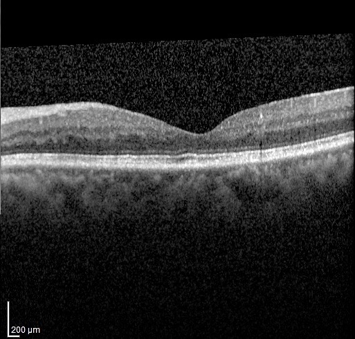
Figure 2. SD-OCT images of the macula showing a right eye macula hole (A) and normal looking macula contour in the left eye (B)

Figure 3. Front view of surgeon and assistant with their 3D glasses on. They are looking at a TV screen instead of peering down the microscope.

Figure 4. View of a large 55’’ 4K monitor screen (Zeiss Artevo 800TM) placed in front of the surgeon, permitting surgery with a more comfortable posture in high resolution. The binoculars of the microscope face upward as they are not used during the 3D surgery.
Reference:
Cheng TC, Yahya MFN, Mohd Naffi AA, Othman O, Seng Fai T, Yong MH, Wan Abdul Halim WH, Mustapha M, Che Hamzah J, Md Din N, Bastion MC. Evaluation of Three-Dimensional Heads up Ophthalmic Surgery Demonstration From the Perspective of Surgeons and Postgraduate Trainees. J Craniofac Surg. 2021 Oct 1;32(7):2285-2291. doi: 10.1097/SCS.0000000000007645. PMID: 33770023.
Reported by: Professor Dr Mae-Lynn Catherine Bastion
Department of Ophthalmology, Faculty of Medicine, UKM & Department of Ophthalmology, Hospital Canselor Tuanku Muhriz