The First Autologous Retinal Transplant (ART) Performed at Hospital Canselor Tuanku Muhriz, UKM
The first autologous retinal transplant (ART) was performed at Hospital Canselor Tuanku Muhriz on Thursday, 7 September 2023 by Professor Dr Mae-Lynn Catherine Bastion, vitreoretinal surgeon and senior consultant ophthalmologist from Department of Ophthalmology, Hospital Canselor Tuanku Muhriz and Faculty of Medicine, Universiti Kebangsaan Malaysia. She is also serves at UKM Specialist Centre. She was assisted by her fellow, Dr Lhacha Wangdi from University of Bhutan, Kingdom of Bhutan. The patient, a 58-year-old retired teacher with history of high myopia (short-sighted power of 18 dioptres) developed left blurred vision and seeing wavy lines (metamorphopsia) after her cataract surgery in January this year at another centre. Our examination found that she had a hole in her left eye macula, responsible for the drop in vision and symptoms. Surgery in the form of vitrectomy and peeling of the internal limiting membrane of the retina with gas bubble injection was performed uneventfully in June 2023. She reported improvement in her vision during her follow up. However, a small persistent hole was detected one month later which rapidly progressed to detachment of her retina (the delicate nerve tissue lining the back of the eye, responsible for vision). Hence she underwent a further left eye vitrectomy, this time with harvest of a small portion of her inferior retina, which was used to repair the defect at the macula (ART) , with the hope that the retina would remain attached after the surgery. She also received a heavy silicone oil bubble this time, allowing her to posture supine after surgery to maintain the graft position. Five days after the operation she is doing well. The picture below shows an optical coherence tomography (OCT) photograph of the graft positioned to cover the macula hole with attached retina surrounding it (Figure 1) and a photograph of the left eye shows the view of the graft (white) at the centre of the macula with the location of the graft site at the lower aspect of the photograph (Figure 2). In 6 weeks’ time, the heavy oil will be removed surgically and we hope she will obtain a good result.
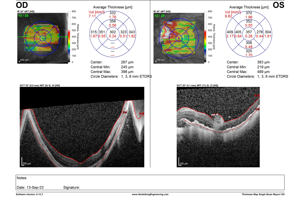
Figure 1. Optical coherence tomography photograph of the ART graft covering the macula hole.
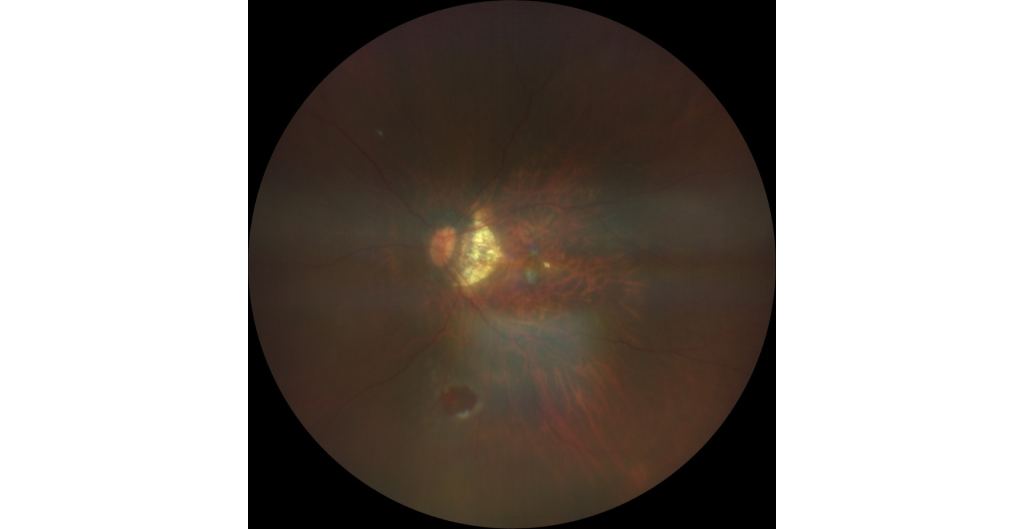
Figure 2. Fundus photograph showing the graft in position at the back of the patient’s eye (covering the macula hole) with the graft taken from the circular area at the bottom of the photo in an oil filled

Surgeons Prof Mae-Lynn Bastion and Dr Lhacha Wangdi, from Department of Ophthalmology, UKM at a dinner function